Bassett Collection of Stereoscopic Images of Human Anatomy
Exploration of the brain from its basal aspect
Inferior occipitofrontal fasciculus, limen insulae and arteries of choroid plexus
Image #5-3
KEYWORDS: Brain, Diencephalon, Telencephalon, Temporal lobe, Vasculature, Ventricules.
Creative Commons
Stanford holds the copyright to the David L. Bassett anatomical images and has assigned Creative Commons license Attribution-Share Alike 4.0 International to all of the images.

For additional information regarding use and permissions, please contact the Medical History Center.

Exploration of the brain from its basal aspect
Inferior occipitofrontal fasciculus, limen insulae and arteries of choroid plexus
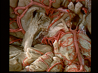
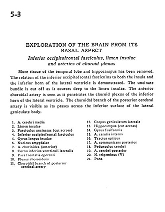
More tissue of the temporal lobe and hippocampus has been removed. The relation of the inferior occipitofrontal fasciculus to both the insula and the inferior horn of the lateral ventricle is demonstrated. The uncinate bundle is cut off as it courses deep to the limen insulae. The anterior choroidal artery is seen as it penetrates the choroid plexus of the inferior horn of the lateral ventricle. The choroidal branch of the posterior cerebral artery is visible as its passes across the inferior surface of the lateral geniculate body.
- Middle cerebral artery
- Limen insulae
- Uncinate fasciculus (cut across)
- Inferior occipitofrontal fasciculus
- Long insular gyrus
- Amygdaloid nucleus
- Choroidal artery (anterior)
- Inferior horn of lateral ventricle
- Frontal part of operculum
- Choroid plexus
- Choroidal branch of posterior cerebral artery
- Lateral geniculate body
- Hippocampus (cut across)
- Fusiform gyrus
- Internal carotid artery
- Optic tract
- Posterior communicating artery
- Cerebral peduncle
- Posterior cerebral artery
- Trigeminal nerve (V)
- Pons