Bassett Collection of Stereoscopic Images of Human Anatomy
General orientation views of dissection
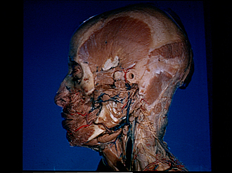
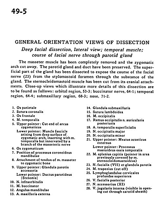
Deep facial dissection, lateral view; temporal muscle; course of facial nerve through parotid gland
Image #49-5
KEYWORDS: Cheek, Connective tissue, Exocrine and endocrine, Face, Muscles and tendons, Peripheral nervous system, Vasculature, Fascia and connective tissue, Overview.
Creative Commons
Stanford holds the copyright to the David L. Bassett anatomical images and has assigned Creative Commons license Attribution-Share Alike 4.0 International to all of the images.

For additional information regarding use and permissions, please contact the Medical History Center.
General orientation views of dissection
Deep facial dissection, lateral view; temporal muscle; course of facial nerve through parotid gland
The masseter muscle has been completely removed and the zygomatic arch cut away. The parotid gland and duct have been preserved. The superficial part of the gland has been dissected to expose the course of the facial nerve (23) from the stylomastoid foramen through the substance of the gland. The sternocleidomastoid muscle has been cut from its cranial attachments. Close-up views which illustrate more details of this dissection are to be found as follows
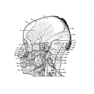
- Parietal bone
- Coronal suture
- Frontal bone
- Temporalis muscle
- Upper pointer: Cut end of zygomatic arch Lower pointer: Muscle tendon arising from deep surface of zygomatic arch, inserting with temporalis muscle but innervated by a branch of the masseteric nerve
- Zygomatic bone
- Position of coronoid process of mandible
- Attachment of tendon of masseter muscle to zygomatic bone
- Upper pointer: Accessory parotid gland Lower pointer: Parotid duct
- Infraorbital nerve
- Buccinator muscle
- Angle of mandible
- External maxillary artery
- Submandibular gland
- Lambdoidal suture
- Occipitalis muscle
- Occipital branch of posterior auricular nerve
- Superficial temporal artery
- Greater occipital nerve
- Lesser occipital nerve
- Upper pointer: External acoustic meatus Lower pointer: Mastoid process
- Splenius capitis muscle (pointer in area previously covered by sternocleidomastoid muscle)
- Facial nerve (VII) and parotid gland
- Trapezius muscle (cut off)
- Superior deep cervical lymph nodes
- Posterior facial vein
- Accessory nerve (XI)
- Internal jugular vein (visible in opening cut through carotid sheath)