
Bassett Collection of Stereoscopic Images of Human Anatomy
Osteology
Deciduous and permanent dentition at six years
Image #47-1
KEYWORDS: Bones cartilage joints, Face, Mouth.
Creative Commons
Stanford holds the copyright to the David L. Bassett anatomical images and has assigned Creative Commons license Attribution-Share Alike 4.0 International to all of the images.

For additional information regarding use and permissions, please contact the Medical History Center.

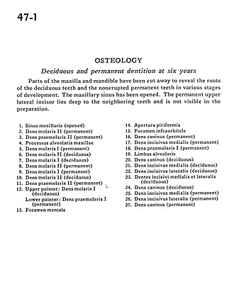
Osteology
Deciduous and permanent dentition at six years
Parts of the maxilla and mandible have been cut away to reveal the roots of the deciduous teeth and the nonerupted permanent teeth in various stages of development. The maxillary sinus has been opened. The permanent upper lateral incisor lies deep to the neighboring teeth and is not visible in the preparation.
- Maxillary sinus (opened)
- Molar II (permanent)
- Premolar II (permanent )
- Alveolar process of maxilla
- Molar I (permanent)
- Molar II (deciduous)
- Molar I (deciduous)
- Molar II (permanent)
- Molar I (permanent)
- Molar II (deciduous)
- Premolar II (permanent)
- Upper pointer: Molar I (deciduous) Lower pointer: Premolar I (permanent)
- Mental foramen
- Piriform aperature
- Infraorbital foramen
- Canine (permanent)
- Medial incisor (permanent)
- Premolar I (permanent)
- Alveolar border
- Canine (deciduous)
- Medial incisor (deciduous)
- Lateral incisor (deciduous)
- Medial and lateral incisors (deciduous)
- Canine (deciduous)
- Medial incisor (permanent)
- Lateral incisor (permanent)
- Canine (permanent)