
Bassett Collection of Stereoscopic Images of Human Anatomy
Osteology
Roentgenogram, left maxilla, mediolateral view
Image #45-3
KEYWORDS: Bones cartilage joints, Face, Mouth.
Creative Commons
Stanford holds the copyright to the David L. Bassett anatomical images and has assigned Creative Commons license Attribution-Share Alike 4.0 International to all of the images.

For additional information regarding use and permissions, please contact the Medical History Center.

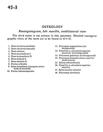
Osteology
Roentgenogram, left maxilla, mediolateral view
The third molar is not present in this specimen. Detailed roentgenographic views of the teeth are to be found in 47-4 ff.
- Medial incisor
- Lateral incisor
- Canine
- Premolar I
- Premolar II
- Molar I
- Molar II
- Orbital surface
- Maxillary sinus (occupies entire body of maxilla)
- Infratemporal surface
- Zygomatic process (in background)
- Canal for superior posterior alveolar artery (in background)
- Palatine process (in foreground)
- Frontal process (note canal for small artery)
- Infraorbital sulcus
- Canal for superior anterior alveolar artery
- Anterior nasal spine
- Alveolar process