
Bassett Collection of Stereoscopic Images of Human Anatomy
Osteology
Lacrimal bones, medial and lateral views
Image #43-7
KEYWORDS: Bones cartilage joints, Face.
Creative Commons
Stanford holds the copyright to the David L. Bassett anatomical images and has assigned Creative Commons license Attribution-Share Alike 4.0 International to all of the images.

For additional information regarding use and permissions, please contact the Medical History Center.
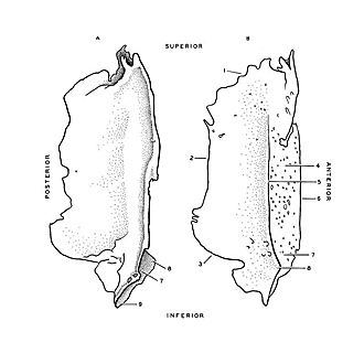

Osteology
Lacrimal bones, medial and lateral views
A. Lacrimal bone left, medial surface.B. Lacrimal bone right, lateral (orbital) surface.
- Margin for articulation with orbital part of frontal bone
- Margin for articulation with lamina papyracea ethmoid bone
- Margin for articulation with maxilla
- Lacrimal sulcus
- Posterior lacrimal crest
- Margin for articulation with frontal process of maxilla
- Fossa of lacrimal sac
- Hamulus of lacrimal bone
- Conchal process for articulation with inferior nasal concha