Bassett Collection of Stereoscopic Images of Human Anatomy
Osteology
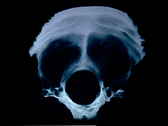
Roentgenogram, occipital bone, internal view
Image #41-4
KEYWORDS: Bones cartilage joints.
Creative Commons
Stanford holds the copyright to the David L. Bassett anatomical images and has assigned Creative Commons license Attribution-Share Alike 4.0 International to all of the images.

For additional information regarding use and permissions, please contact the Medical History Center.

Osteology
Roentgenogram, occipital bone, internal view
Numerous channels for diploic veins (4) are visible close to the dense bone which bounds the foramen magnum (8) posteriorly and laterally. These channels are continuous upward into the squamous part of the bone near the midline. Less prominent bilateral channels are present near the mastoid border of the bone.
- Squamous part of occipital bone
- Lambdoidal margin
- Transverse sulcus
- Channels within bone for occipital diploic veins
- Mastoid margin
- Occipital condyle
- Basilar part
- Foramen magnum
- Jugular tubercle
- Jugular incisure
- Hypoglossal canal