Bassett Collection of Stereoscopic Images of Human Anatomy
Osteology
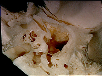
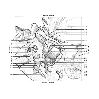
Left temporal bone, inferior view; middle and inner ear cavities dissected
Image #41-1
KEYWORDS: Bones cartilage joints, Ear.
Creative Commons
Stanford holds the copyright to the David L. Bassett anatomical images and has assigned Creative Commons license Attribution-Share Alike 4.0 International to all of the images.
For additional information regarding use and permissions, please contact the Medical History Center.
Osteology
Left temporal bone, inferior view; middle and inner ear cavities dissected
The petrous and tympanic parts of the bone have been ground away from below to expose the tympanic cavity (middle ear), which occupies the central area of the view, and the osseous labyrinth (inner ear), medial to the tympanic cavity. The cochlea has been opened parallel to the modiolus (6) to expose its basal, middle, and apical turns.
- Carotid canal
- Helicotrema
- Wall of modiolus
- Upper pointer: Scala vestibuli Lower pointer: Scala tympani
- Spiral canal of modiolus
- Base of modiolus
- Internal acoustic meatus (pointer on transverse crest)
- Lamina bony spiral
- Spherical recess of vestibule (rough white area in view is macula cribrosa superior)
- Upper pointer: Superior bony ampulla (deep in vestibule) Lower pointer: Eliptical recess of vestibule
- Opening of common crus
- Upper pointer: posterior bony ampulla Lower pointer: Posterior semicircular canal
- Mandibular fossa
- Semicanal of auditory tube
- Tensor tympani muscle
- Petrotympanic fissure (pointer indicates site of emergence of chorda tympani)
- Cut edge of tympanic part
- Cochleariform process
- Epitympanic recess (deep part of cavity)
- Position of oval window (obscured by promontorium)
- External acoustic meatus
- Prominence of facial canal
- Promontorium (note promontory sulcus for tympanic plexus)
- Round window (cut open)
- Pyramidal eminence
- Tympanic sinus
- Canal for stapedius muscle
- Facial canal


