Bassett Collection of Stereoscopic Images of Human Anatomy
Osteology
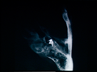
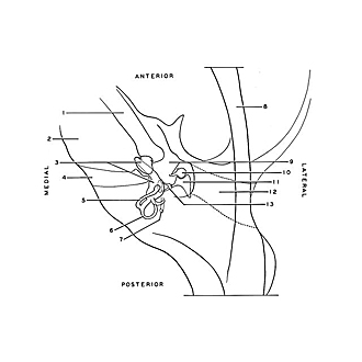
Roentgenogram, right temporal bone; ear ossicles in situ
Image #40-6
KEYWORDS: Bones cartilage joints, Ear.
Creative Commons
Stanford holds the copyright to the David L. Bassett anatomical images and has assigned Creative Commons license Attribution-Share Alike 4.0 International to all of the images.

For additional information regarding use and permissions, please contact the Medical History Center.

Osteology
Roentgenogram, right temporal bone; ear ossicles in situ
The temporal bone was decalcified after removal of the ear ossicles. These ossicles were then replaced in their normal positions. The view is from above. The ossicles are shown in detail in 62-6 ff.
- Carotid canal
- Petrosal part
- Upper pointer: Cochlea Lower pointer: Vestibulum
- Internal acoustic meatus
- Semicircular canal superior
- Semicircular canal posterior
- Stylomastoid foramen
- Temporal bone (squamous part)
- Upper pointer: Tympanic cave Lower pointer: Manubrium of malleus
- Capitulum of malleus
- Body incus
- External acoustic meatus
- Stapes (base of stapes lies in fenestra vestibuli medial to pointer)