
Bassett Collection of Stereoscopic Images of Human Anatomy
Osteology
Roentgenogram, right temporal bone, superior view
Image #40-5
KEYWORDS: Bones cartilage joints, Ear.
Creative Commons
Stanford holds the copyright to the David L. Bassett anatomical images and has assigned Creative Commons license Attribution-Share Alike 4.0 International to all of the images.

For additional information regarding use and permissions, please contact the Medical History Center.
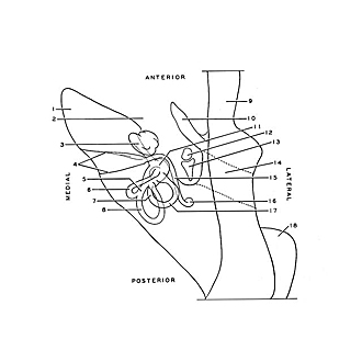

Osteology
Roentgenogram, right temporal bone, superior view
The contours of the bony labyrinth are visible because of the air which fills the labyrinth and contrasts with the surrounding petrous bone.
- Petrosal part
- Carotid canal (partially beneath cochlea)
- Cochlea
- Borders of internal acoustic meatus
- Common crus semicircular canal
- Semicircular canal superior (note ampulla near junction of anterior crus with vestibule)
- Crus simplex semicircular canal
- Semicircular canal posterior (ampulla is visible at inferior termination of canal in vestibule)
- Temporal bone (squamous part)
- Tympanic part
- Vestibulum
- Malleus (faintly visible within middle ear cavity)
- Incus (faintly visible)
- External acoustic meatus (beneath squama)
- Facial canal
- Stylomastoid foramen (in background)
- Semicircular canal lateral (ampulla difficult to distinguish at anterior termination of canal)
- Mastoid process