
Bassett Collection of Stereoscopic Images of Human Anatomy
Osteology
Left temporal bone, superior view
Image #40-4
KEYWORDS: Bones cartilage joints.
Creative Commons
Stanford holds the copyright to the David L. Bassett anatomical images and has assigned Creative Commons license Attribution-Share Alike 4.0 International to all of the images.

For additional information regarding use and permissions, please contact the Medical History Center.

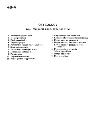
Osteology
Left temporal bone, superior view
- Zygomatic process
- Parietal margin
- Cerebral surface
- Tegmen tympani
- Remnant of petrosquamous fissure
- Temporal bone (squamous part)
- Sulcus for middle meningeal artery
- Hiatus facial canal
- Petrosal part
- Trigeminal impression
- Posterior pyramidal surface
- Superior pyramidal angle
- Location of internal acoustic meatus
- Anterior pyramidal surface
- Upper pointer: Arcuate eminence Lower pointer: Superior petrosal sulcus
- Intrajugular process
- Sigmoid sulcus
- Occipital margin
- Mastoid part