Bassett Collection of Stereoscopic Images of Human Anatomy
Osteology
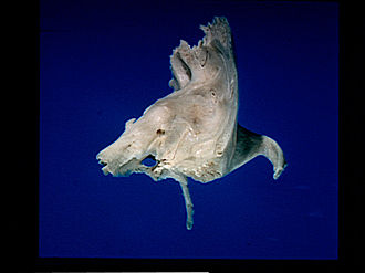
Left temporal bone, anterior view
Image #40-2
KEYWORDS: Bones cartilage joints.
Creative Commons
Stanford holds the copyright to the David L. Bassett anatomical images and has assigned Creative Commons license Attribution-Share Alike 4.0 International to all of the images.

For additional information regarding use and permissions, please contact the Medical History Center.
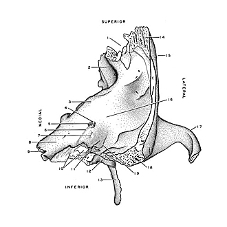
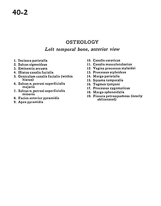
Osteology
Left temporal bone, anterior view
- Parietal incisure
- Sigmoid sulcus
- Arcuate eminence
- Hiatus facial canal
- Geniculum of facial canal (within hiatus)
- Sulcus greater (superficial) petrosal nerve
- Sulcus lesser petrosal (superficial) nerve
- Anterior pyramidal surface
- Pyramidal apex
- Carotid canal
- Canal for Tensor tympani
- Sheath of styloid process
- Styloid process
- Parietal margin
- Temporal bone (squamous part)
- Tegmen tympani
- Zygomatic process
- Sphenoidal margin
- Petrosquamous fissure (nearly obliterated)