Bassett Collection of Stereoscopic Images of Human Anatomy
Osteology
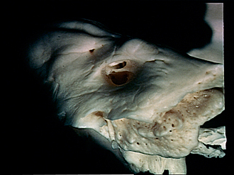
Left temporal bone; close-up view of fundus of internal auditory meatus
Image #40-1
KEYWORDS: Bones cartilage joints, Ear.
Creative Commons
Stanford holds the copyright to the David L. Bassett anatomical images and has assigned Creative Commons license Attribution-Share Alike 4.0 International to all of the images.

For additional information regarding use and permissions, please contact the Medical History Center.


Osteology
Left temporal bone; close-up view of fundus of internal auditory meatus
The crista transversa (11) divides the fundus into a superior and inferior fossa. The apex of the petrous pyramid is to the right of the view.
- Superior petrous sulcus
- Subarcuate fossa
- Superior vestibular area
- Inferior vestibular area
- Foramen singulare (faintly visible)
- External opening of vestibular aqueduct
- External opening cochlear canal
- Arcuate eminence
- Superior pyramidal angle
- Area facial nerve
- Crista transversa
- Cochlear area
- Margin of internal acoustic meatus
- Posterior pyramidal angle
- Inferior pyramidal surface