Bassett Collection of Stereoscopic Images of Human Anatomy
Exploration of the brain from its basal aspect
Relations of tip of inferior horn of lateral ventricle
Image #4-5
KEYWORDS: Brain, Meninges, Telencephalon, Temporal lobe, Ventricules.
Creative Commons
Stanford holds the copyright to the David L. Bassett anatomical images and has assigned Creative Commons license Attribution-Share Alike 4.0 International to all of the images.

For additional information regarding use and permissions, please contact the Medical History Center.
Exploration of the brain from its basal aspect
Relations of tip of inferior horn of lateral ventricle
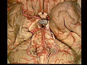
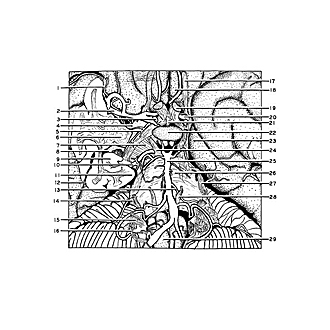
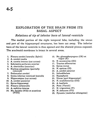
The medial portion of the right temporal lobe, including the uncus and part of the hippocampal structures, has been cut away. The inferior horn of the lateral ventricle is thus opened and the choroid plexus exposed. The arachnoid membrane is intact in several areas.
- Lateral cerebral fissure (Sylvian)
- Middle cerebral artery
- Internal carotid artery (cut across)
- Anterior perforated substance
- Choroidal artery (anterior)
- Amygdaloid nucleus (partially removed)
- Cerebral peduncle
- Inferior horn of lateral ventricle
- Hippocampus (cut across)
- Posterior cerebral artery
- Hippocampal gyrus
- Collateral fissure
- Internal auditory artery
- Facial nerve (VII) and vestibulocochlear (VIII)
- Glossopharyngeal (IX) and vagus (X) nerves
- Accessory nerve (XI)
- Olfactory tract
- Straight gyrus
- Optic nerve (II)
- Anterior cerebral artery
- Infundibulum
- Pituitary
- Uncus (hippocampal gyrus)
- Oculomotor nerve (III)
- Basilar artery
- Pons
- Trigeminal nerve (V)
- Abducens nerve (VI)
- Medulla oblongata (pyramid)