Bassett Collection of Stereoscopic Images of Human Anatomy
Creative Commons
Stanford holds the copyright to the David L. Bassett anatomical images and has assigned Creative Commons license Attribution-Share Alike 4.0 International to all of the images.

For additional information regarding use and permissions, please contact the Medical History Center.

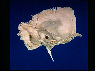
Osteology
Left temporal bone, medial view
The petrous part of the bone projects into the foreground. The squamous part lies above and to its right, the mastoid portion to the left.
- Parietal margin
- Parietal incisura
- Sigmoid sulcus
- Mastoid foramen
- Occipital margin
- Superior petrous sulcus & subarcuate fossa
- Aperture of external vestibular aqueduct
- Posterior pyramidal surface
- External aperture cochlear canaliculi
- Internal acoustic meatus
- Posterior pyramidal angle
- Inferior pyramidal surface
- Temporal bone (squamous part)
- Cerebral surface
- Groove for posterior branch of middle meningeal artery
- Arcuate eminence
- Tegmen tympani
- Zygomatic process
- Sphenoidal margin
- Ant. pyramidal surface
- Pyramidal apex
- Styloid process