Bassett Collection of Stereoscopic Images of Human Anatomy
Osteology
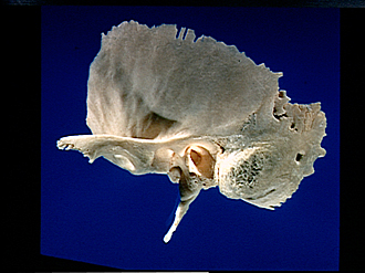
Left temporal bone, lateral view
Image #39-5
KEYWORDS: Bones cartilage joints.
Creative Commons
Stanford holds the copyright to the David L. Bassett anatomical images and has assigned Creative Commons license Attribution-Share Alike 4.0 International to all of the images.

For additional information regarding use and permissions, please contact the Medical History Center.


Osteology
Left temporal bone, lateral view
- Temporal bone (squamous part)
- Temporal surface
- Posterior root of zygomatic process
- Zygomatic process
- Anterior root of zygomatic process and articular tubercle
- Mandibular fossa
- Middle root of zygomatic process
- Petrotympanic fissure
- Tympanic part
- Sheath of styloid process
- Styloid process
- Parietal margin
- Parietal incisura (note sutural bone)
- Temporal line
- Sulcus middle temporal artery
- Suprameatal spine and mastoid fossa
- Mastoid foramen
- Tympanomastoid fissure
- Mastoid part
- Sulcus for occipital artery
- Mastoid incisura
- Mastoid process
- External acoustic meatus