
Bassett Collection of Stereoscopic Images of Human Anatomy
Osteology
Right temporal, infratemporal and pterygopalatine fossae, inferolateral view
Image #38-5
KEYWORDS: Bones cartilage joints.
Creative Commons
Stanford holds the copyright to the David L. Bassett anatomical images and has assigned Creative Commons license Attribution-Share Alike 4.0 International to all of the images.

For additional information regarding use and permissions, please contact the Medical History Center.
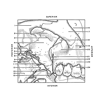
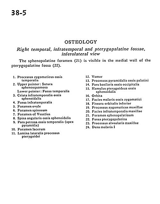
Osteology
Right temporal, infratemporal and pterygopalatine fossae, inferolateral view
The sphenopalatine foramen (21) is visible in the medial wall of the pterygopalatine fossa (22).
- Zygomatic process temporal bone
- Upper pointer: Sphenosquamous suture Lower pointer: Temporal fossa
- Infratemporal crest sphenoid bone
- Infratemporal fossa
- Foramen ovale
- Foramen spinosum
- Foramen of Vesalius
- Angular spine sphenoid bone
- Petrosal part of temporal bone (apex pyramidis)
- Foramen lacerum
- Lateral plate of pterygoid process
- Vomer
- Pyramidal process palatine bone
- Basal part occipital bone
- Pterygoid hàmulus sphenoid bone
- Orbit
- Surface malaris zygomatic bone
- Inferior orbital fissure
- Zygomatic process of maxilla
- Infratemporal surface of maxilla
- Sphenopalatine foramen
- Pterygopalatine fossa
- Alveolar process of maxilla
- First molar