Bassett Collection of Stereoscopic Images of Human Anatomy
Osteology
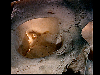
Right orbit, anterior view
Image #37-1
KEYWORDS: Bones cartilage joints, Eye, Face.
Creative Commons
Stanford holds the copyright to the David L. Bassett anatomical images and has assigned Creative Commons license Attribution-Share Alike 4.0 International to all of the images.

For additional information regarding use and permissions, please contact the Medical History Center.


Osteology
Right orbit, anterior view
- Supraorbital margin
- Orbital surface of frontal bone
- Sphenofrontal suture
- Zygomaticofrontal suture
- Superior orbital fissure
- Upper pointer: Optic foramen Lower pointer: Lateral rectus spine
- Lower root of Lesser wing sphenoid bone
- Upper pointer: Orbital tubercle of zygomatic bone Lower pointer: Body sphenoid bone
- Orbital process palatine bone
- Greater wing of sphenoid
- Sphenozygomatic suture
- Inferior orbital fissure
- Zygomatic bone
- Upper pointer: Ethmoidomaxillary suture Lower pointer: Orbital surface of maxilla
- Infraorbital sulcus
- Zygomaticomaxillary suture
- Supraorbital foramen
- Nasal part of frontal bone
- Anterior ethmoidal foramen
- Nasofrontal suture
- Frontomaxillary suture
- Posterior ethmoidal foramen
- Upper pointer: Orbital plate of ethmoid Lower pointer: Lacrimoethmoidal suture
- Lacrimomaxillary suture
- Fossa of lacrimal sac
- Anterior lacrimal crest
- Posterior lacrimal crest
- Frontal process of maxilla
- Infraorbital margin
- Upper pointer: Infraorbital suture Lower pointer: Infraorbital foramen