Bassett Collection of Stereoscopic Images of Human Anatomy
Osteology
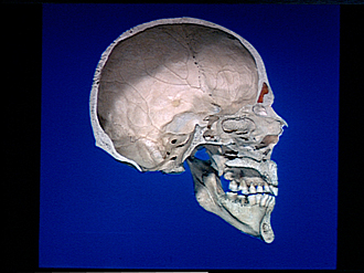
Skull, midsagittal section
Image #36-4
KEYWORDS: Bones cartilage joints, Face, Mouth, Overview.
Creative Commons
Stanford holds the copyright to the David L. Bassett anatomical images and has assigned Creative Commons license Attribution-Share Alike 4.0 International to all of the images.

For additional information regarding use and permissions, please contact the Medical History Center.


Osteology
Skull, midsagittal section
- Parietal bone
- Greater wing of sphenoid & sphenoparietal suture
- Squamous suture &temporal bone (squamous part)
- Occipital bone (squamous part)
- Lambdoidal suture
- Sulcus for posterior branch of middle meningeal artery
- Petrosal part of temporal bone & internal acoustic meatus
- Occipitomastoid suture
- Sigmoid sulcus
- Foramen magnum (lateral margin)
- Hypoglossal canal
- Occipital condyle
- Basi occiput
- Pterygoid process sphenoid bone and choana
- Mandibular foramen
- Horizontal plate palatine bone
- Mylohyoid sulcus
- Mandible
- Mylohyoid line
- Coronal suture
- Frontal bone (squamous part)
- Sulcus for anterior branch of middle meningeal artery
- Frontal sinus
- Nasion
- Nasal bone
- Crista gall & lamina cribrosa
- Superior nasal concha (visible through opening in nasal septum)
- Perpendicular plate of ethmoid bone (partially cut away posteriorly)
- Sphenoid sinus
- Vomer
- Inferior nasal concha
- Nasal incisura
- Palatine process of maxilla
- Alveolar process of maxilla