Bassett Collection of Stereoscopic Images of Human Anatomy
Osteology
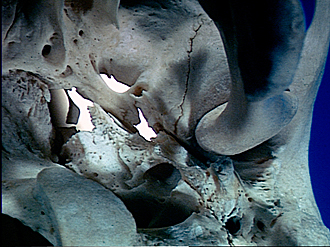
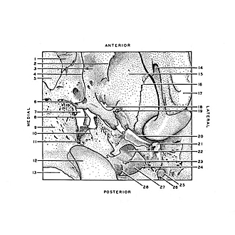
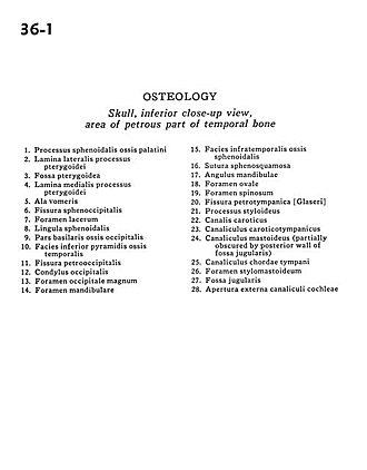
Skull, inferior close-up view, area of petrous part of temporal bone
Image #36-1
KEYWORDS: Bones cartilage joints.
Creative Commons
Stanford holds the copyright to the David L. Bassett anatomical images and has assigned Creative Commons license Attribution-Share Alike 4.0 International to all of the images.

For additional information regarding use and permissions, please contact the Medical History Center.
Osteology
Skull, inferior close-up view, area of petrous part of temporal bone
- Sphenoid process palatine bone
- Lateral plate of pterygoid process
- Pterygoid fossa
- Medial plate of pterygoid process
- Ala of vomer
- Sphenooccipital fissure
- Foramen lacerum
- Lingula of sphenoid
- Basal part occipital bone
- Inferior surface of temporal bone
- Petro-occipital fissure
- Occipital condyle
- Foramen magnum
- Mandibular foramen
- Infratemporal surface of sphenoid bone
- Sphenosquamous suture
- Angle of mandible
- Foramen ovale
- Foramen spinosum
- Petrotympanic fissure
- Styloid process
- Carotid canal
- Tympanic canaliculus
- Mastoid canaliculus (partially obscured by posterior wall of jugular fossa)
- Canaliculus chordae tympani
- Stylomastoid foramen
- Jugular fossa
- External aperture of cochlear canaliculi