Bassett Collection of Stereoscopic Images of Human Anatomy
Osteology
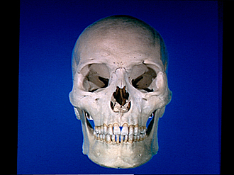
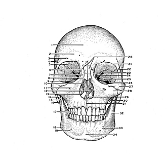
Skull, anterior view
Image #35-4
KEYWORDS: Bones cartilage joints, Cheek, Eye, Face, Mouth, Nose, Overview.
Creative Commons
Stanford holds the copyright to the David L. Bassett anatomical images and has assigned Creative Commons license Attribution-Share Alike 4.0 International to all of the images.

For additional information regarding use and permissions, please contact the Medical History Center.

Osteology
Skull, anterior view
A supraorbital foramen occurs on the right, a supraorbital notch on the left. Note that within the notch there is a small superciliary canal which extends into the frontal bone. This canal gives passage to a small nutrient artery, an emissary vein, and a nerve filament. The deviation of the bony nasal septum is not unusual.
- Frontal bone (squamous part)
- Superciliary arch
- Supraorbital foramen
- Temporal fossa
- Orbital surface of frontal bone
- Frontomaxillary suture
- Frontal process of maxilla
- Nasomaxillary suture
- Orbital surface of maxilla
- Middle nasal concha
- Infraorbital foramen
- Bony nasal septum
- Inferior nasal concha
- Neck of condylar process of mandible
- Coronoid process of mandible
- Mastoid process
- Canine
- Angle of mandible
- Mental foramen
- Nasofrontal suture & Nasal bone
- Frontal incisura
- Zygomaticofrontal suture
- Superior orbital fissure
- Optic foramen
- Orbital surface of the greater wing of the sphenoid and Sphenozygomatic suture
- Inferior orbital fissure
- Zygomatic bone
- Zygomaticomaxillary suture & Zygomatic process of maxilla
- Anterior nasal spine
- Styloid process (temporal bone)
- Intermaxillary suture
- Ramus of mandible
- Body of mandible
- Mental protuberance