
Bassett Collection of Stereoscopic Images of Human Anatomy
Osteology
Roentgenogram of skull, left lateral view
Image #35-2
KEYWORDS: Bones cartilage joints, Overview.
Creative Commons
Stanford holds the copyright to the David L. Bassett anatomical images and has assigned Creative Commons license Attribution-Share Alike 4.0 International to all of the images.

For additional information regarding use and permissions, please contact the Medical History Center.

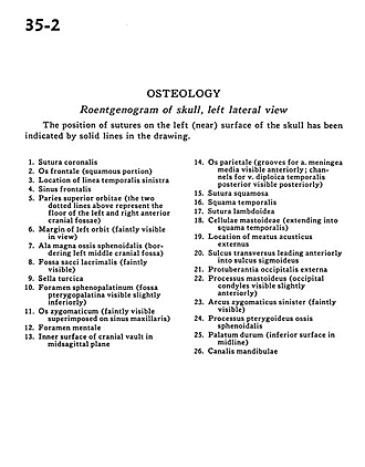
Osteology
Roentgenogram of skull, left lateral view
The position of sutures on the left (near) surface of skull has been indicated by solid lines in the drawing.
- Coronal suture
- Frontal bone (squamous portion)
- Location of temporal line left
- Frontal sinus
- Superior wall of orbit (the two dotted lines above represent the floor of the left and right anterior cranial fossae) 6. Margin of left orbit (faintly visible in view)
- Greater wing of sphenoid (bordering left middle cranial fossa)
- Fossa of lacrimal sac (faintly visible)
- Sella turcica
- Sphenopalatine foramen (Pterygopalatine fossa visible slightly inferiorly)
- Zygomatic bone (faintly visible superimposed on maxillary sinus)
- Mental foramen
- Inner surface of cranial vault in midsagittal plane
- Parietal bone (grooves for middle meningeal artery visible anteriorly; channels for temporal posterior diploic vein visible posteriorly)
- Squamous suture
- Temporal bone (squamous part)
- Lambdoidal suture
- Mastoid cells (extending into temporal bone (squamous part)
- Location of external acoustic meatus
- Transverse sinus leading anteriorly into sigmoid sinus
- External occipital protuberance
- Mastoid process (occipital condyles visible slightly anteriorly)
- Zygomatic arch left (faintly visible)
- Pterygoid process of sphenoid bone
- Hard palate (inferior surface in midline)
- Mandibular canal