Bassett Collection of Stereoscopic Images of Human Anatomy
Osteology
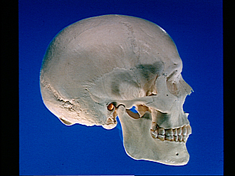
Skull, right lateral view
Image #35-1
KEYWORDS: Bones cartilage joints, Overview.
Creative Commons
Stanford holds the copyright to the David L. Bassett anatomical images and has assigned Creative Commons license Attribution-Share Alike 4.0 International to all of the images.

For additional information regarding use and permissions, please contact the Medical History Center.


Osteology
Skull, right lateral view
- Parietal bone
- Superior temporal line
- Inferior temporal line
- Squamous suture
- Sphenosquamous suture
- Temporal bone (squamous part)
- Lambdoidal suture
- Occipital bone (squamous part)
- Intersutural bone
- Occipitomastoid suture
- External occipital protuberance (unusually prominent)
- Upper pointer: External acoustic meatus (tympanic prominence visible in interior) Lower pointer: Condylar process of mandible
- Mastoid process
- Styloid process
- Occipital condyle
- Lateral plate of pterygoid process
- Upper pointer: Coronoid process of mandible Lower pointer: Ramus of mandible
- Coronal suture
- Frontal bone (squamous part)
- Sphenoparietal suture
- Sphenofrontal suture
- Greater wing of sphenoid
- Zygomaticofrontal suture
- Frontolacrimal suture
- Lacrimomaxillary suture
- Fossa of lacrimal sac
- Nasomaxillary suture
- Sphenozygomatic suture
- Zygomatic arch (pointer indicates zygomaticotemporal suture)
- Zygomaticomaxillary suture
- Zygomatic bone
- Anterior nasal spine
- First molar
- Body of mandible (pointer near mental foramen)