Bassett Collection of Stereoscopic Images of Human Anatomy
Exploration of the spinal cord and meninges in situ
Roots and ganglion of 10th thoracic nerve
Image #33-4
KEYWORDS: Central nervous system, Thoracic region, Vertebral column.
Creative Commons
Stanford holds the copyright to the David L. Bassett anatomical images and has assigned Creative Commons license Attribution-Share Alike 4.0 International to all of the images.

For additional information regarding use and permissions, please contact the Medical History Center.

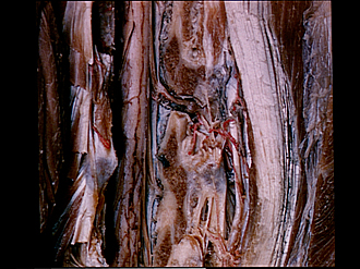
Exploration of the spinal cord and meninges in situ
Roots and ganglion of 10th thoracic nerve
The articular processes of the tenth and eleventh thoracic vertebrae have been cut away on the right side to reveal the lateral course of the 10th thoracic spinal roots. Subarachnoid spaces extend only a millimeter or two along the anterior and posterior roots after they pierce the dura mater. A narrow bridge of dura intervenes between the motor and sensory roots. A mass of plexiform veins was removed from the intervertebral foramen.
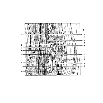
- Thoracic part spinal cord
- Internal vertebral venous plexus
- Superior articular surface thoracic vertebra XI
- External posterior spinal vein
- Vein within arch of 11th thoracic vertebra
- Levator costarum muscle
- Ligamentum flavum
- Superior articular process thoracic vertebra XII
- Superior articular process thoracic vertebra X
- Dura mater
- Dorsal root thoracic nerve X
- Spinal branch of intercostal artery (anterior root of nerve lies just above this)
- Spinal ganglion thoracic nerve X
- Posterior branch of thoracic nerve X
- Transverse process thoracic vertebra XI
- Longissimus dorsi muscle
- Ligamentum intertransversarium
- Inferior articular process thoracic vertebra XI