Bassett Collection of Stereoscopic Images of Human Anatomy
Serial transverse sections of the brain stem
Diencephalon and telencephalon; level of anterior commissure
Image #31-5
KEYWORDS: Brain, Diencephalon, Telencephalon, Ventricules.
Creative Commons
Stanford holds the copyright to the David L. Bassett anatomical images and has assigned Creative Commons license Attribution-Share Alike 4.0 International to all of the images.

For additional information regarding use and permissions, please contact the Medical History Center.
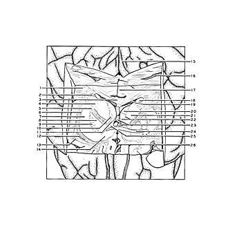
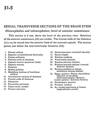
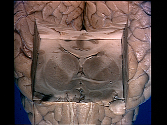
Serial transverse sections of the brain stem
Diencephalon and telencephalon; level of anterior commissure
This section is 6 mm. above the level of the previous view. Relations of the anterior commissure (24) are visible. The frontal stalk of the thalamus (11) can be traced into the anterior limb of the internal capsule. The section passes just below the interventricular foramina (23).
- Corona radiata
- Superior occipitofrontal fasciculus
- Corpus callosum
- Parietal stalk of thalamus
- Internal capsule (posterior limb)
- External capsule
- Putamen
- Globus pallidus
- Medullary laminae of lentiform nucleus
- Intralaminar nucleus of thalamus
- Frontal stalk of thalamus
- Claustrum
- Uncinate fasciculus
- Head of caudate nucleus
- Calcarine fissure
- Posterior horn lateral ventricle
- Cingulate gyrus
- Caudate nucleus
- Lateral ventricle
- Anterior nucleus of thalamus
- Lateral nucleus of thalamus (ventral anterior nucleus)
- Medial nucleus and internal medullary lamina of thalamus
- Choroid plexus adjacent to interventricular foramen (of Monro)
- Anterior commissure
- Preoptic nucleus
- Anterior cerebral arteries and longitudinal fissure (cerebral)