Bassett Collection of Stereoscopic Images of Human Anatomy
Serial transverse sections of the brain stem
Diencephalon and telencephalon.
Image #31-4
KEYWORDS: Brain, Diencephalon, Telencephalon.
Creative Commons
Stanford holds the copyright to the David L. Bassett anatomical images and has assigned Creative Commons license Attribution-Share Alike 4.0 International to all of the images.

For additional information regarding use and permissions, please contact the Medical History Center.

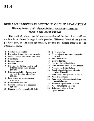
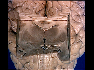
Serial transverse sections of the brain stem
Diencephalon and telencephalon.
The level of this section is 7 mm. above that of the last. The lentiform nucleus is sectioned through its mid-portion. Efferent fibers of the globus pallidus pass, as the ansa lenticularis, around the medial margin of the internal capsule.
- Caudate nucleus (tail)
- Posterior limb of internal capsule
- Dorsal lateral nucleus of thalamus
- Claustrum
- External capsule
- Putamen
- External medullary lamina and external division of globus pallidus
- Internal medullary lamina and internal division of globus pallidus
- Posterior part of anterior commissure
- Uncinate fasciculus
- Lamina terminalis and optic recess
- Lateral cerebral fissure
- Orbital gyri
- Cingulate gyrus and sulcus corpus callosum
- Stria terminalis
- Fornix (body)
- Lateral nucleus of thalamus
- Internal medullary lamina of thalamus
- Medial nucleus of thalamus
- Mamillothalamic fasciculus [Vicq d'Azyr]
- Frontal part internal capsule
- Ansa lenticularis
- Tectal part of column of fornix and hypothalamus
- Anterior perforated substance
- Anterior communicating artery
- Olfactory trigone
- Straight gyrus