Bassett Collection of Stereoscopic Images of Human Anatomy
Serial transverse sections of the brain stem
Pons.
Image #30-5
KEYWORDS: Brain, Cerebellum, Peripheral nervous system, Pons.
Creative Commons
Stanford holds the copyright to the David L. Bassett anatomical images and has assigned Creative Commons license Attribution-Share Alike 4.0 International to all of the images.
For additional information regarding use and permissions, please contact the Medical History Center.
Serial transverse sections of the brain stem
Pons.
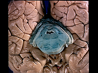
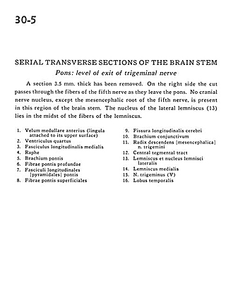
A section 3.5 mm. thick has been removed. On the right side the cut passes through the fibers of the fifth nerve as they leave the pons. No cranial nerve nucleus, except the mesencephalic root of the fifth nerve, is present in this region of the brain stem. The nucleus of the lateral lemniscus (13) lies in the midst of the fibers of the lemniscus.
- Anterior medullary velum (lingula attached to its upper surface)
- Fourth ventricle
- Medial longitudinal fasciculus
- Raphe
- Brachium pontis (middle cerebellar peduncle)
- Deep pontine fibers
- Longitudinal fasciculus
- Superficial pontine fibers
- Longitudinal fissure (cerebral)
- Brachium conjunctivum (superior cerebellar peduncle)
- Descending root (mesencephalic) of trigeminal nerve (V)
- Central tegmental tract
- Lateral lemniscus and nucleus
- Medial lemniscus
- Trigeminal nerve (V)
- Temporal lobe


