
Bassett Collection of Stereoscopic Images of Human Anatomy
Exploration of the meninges and brain in situ
Brain stem and cranial nerves
Image #3-6
KEYWORDS: Brain, Midbrain, Peripheral nervous system.
Creative Commons
Stanford holds the copyright to the David L. Bassett anatomical images and has assigned Creative Commons license Attribution-Share Alike 4.0 International to all of the images.

For additional information regarding use and permissions, please contact the Medical History Center.

Exploration of the meninges and brain in situ
Brain stem and cranial nerves
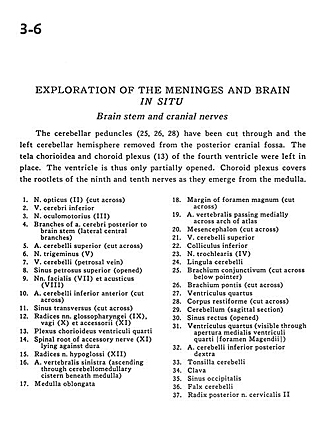
The cerebellar peduncles (25, 26, 28) have been cut through and the left cerebellar hemisphere removed from the posterior cranial fossa. The tela chorioidea and choroid plexus (13) of the fourth ventricle were left in place. The ventricle is thus only partially opened. Choroid plexus covers the rootlets of the ninth and tenth nerves as they emerge from the medulla.
- Optic nerve (II) (cut across)
- Inferior cerebral vein
- Oculomotor nerve (III)
- Branches of cerebral artery posterior to brain stem (lateral central branches)
- Superior cerebellar artery (cut across)
- Trigeminal nerve (V)
- Cerebellar vein (petrosal vein)
- Superior petrosal sinus (opened)
- Facial nerve (VII) and vestibulocochlear nerve (VIII)
- Anterior inferior cerebellar artery (cut across)
- Transverse sinus (cut across)
- Glossopharyngeal nerve (IX), vagus nerve (X), and accessory nerve (Xl)
- Choroid plexus fourth ventricle
- Spinal root of accessory nerve (XI) lying against dura
- Roots hypoglossal nerve (XII)
- Verterbral artery left (ascending through cerebellomedullary cistern beneath medulla)
- Medulla oblongata
- Margin of foramen magnum (cut across)
- Vertebral artery passing medially across arch of atlas
- Mesencephalon (cut across)
- Superior cerebellar vein
- Inferior colliculus
- Trochlear nerve (IV)
- Cerebellar lingula
- Superior cerebellar peduncle (cut across below pointer)
- Brachium pontis (middle cerebellar peduncle) (cut across)
- Fourth ventricle
- Inferior cerebellar peduncle(cut across)
- Cerebellum (sagittal section)
- Straight sinus (opened)
- Fourth ventricle (visible through medial aperture fourth ventricle [foramen of Magendie])
- Posterior inferior cerebellar artery right
- Cerebellar tonsil
- Clava
- Occipital sinus
- Falx cerebelli
- Root of cervical nerve (II)