Bassett Collection of Stereoscopic Images of Human Anatomy
Exploration of the basal aspects of the medulla, pons and cerebellum
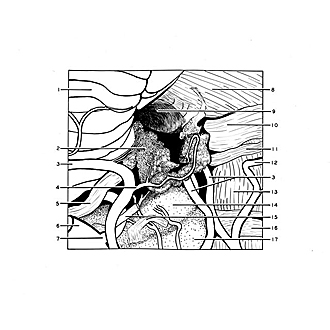
Arteries supplying choroid plexus of lateral recess
Image #28-5
KEYWORDS: Brain, Cerebellum, Medulla, Vasculature, Ventricules.
Creative Commons
Stanford holds the copyright to the David L. Bassett anatomical images and has assigned Creative Commons license Attribution-Share Alike 4.0 International to all of the images.
For additional information regarding use and permissions, please contact the Medical History Center.
Exploration of the basal aspects of the medulla, pons and cerebellum
Arteries supplying choroid plexus of lateral recess
This view is at right angles to the two previous ones, and the midline of the brain stem is now at the bottom of the view. The flocculus has been removed and the anterior inferior cerebellar artery turned medially together with the choroid plexus previously seen. One principal choroidal artery and several small vessels are thus exposed in their course into the plexus. In the depths of the dissection the peduncle of the flocculus is visible. The bulbar roots of Nn. IX, X, and XI have been cut away.
- Tonsil (ventral paraflocculus)
- Choroid plexus fourth ventricle
- Anterior inferior cerebellar artery
- Choroidal artery
- Accessory nerve (XI)
- Vertebral artery right
- Point of origin of posterior inferior cerebellar artery
- Brachium pontis (middle cerebellar peduncle)
- Floccular peduncle
- Vestibulocochlear nerve (VIII)
- Facial nerve (VII)
- Internal auditory artery
- Pons
- Olive
- Hypoglossal nerve (XII)
- Abducens nerve (VI)
- Pyramid (medulla oblongata)


