Bassett Collection of Stereoscopic Images of Human Anatomy
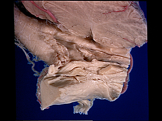
Exploration of the cerebellum and brain stem from above and to the right
Cerebellar peduncles, rhomboid fossa, peduncle of flocculus and flocculotegmental fibers
Image #27-7
KEYWORDS: Brain, Cerebellum, Midbrain, Pons, Ventricules.
Creative Commons
Stanford holds the copyright to the David L. Bassett anatomical images and has assigned Creative Commons license Attribution-Share Alike 4.0 International to all of the images.
For additional information regarding use and permissions, please contact the Medical History Center.
Exploration of the cerebellum and brain stem from above and to the right
Cerebellar peduncles, rhomboid fossa, peduncle of flocculus and flocculotegmental fibers
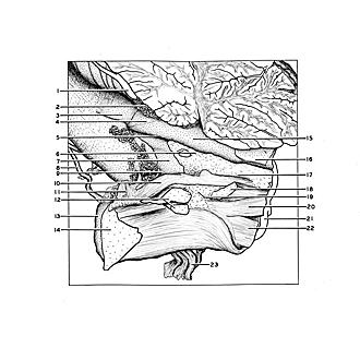
The right cerebellar hemisphere has been cut away so that the relations of its three peduncles are visible. The rhomboid fossa, which forms the floor of the fourth ventricle, has been widely exposed. The ependymal lining of the floor of the ventricle has been cut away except for a small island over the facial colliculus (6). The pigmentation of the locus caeruleus (17) is visible as it extends toward the mesencephalon. The peduncle of the flocculus (11) projects upward from behind the brachium pontis and is broken off. Fibers diverging from the peduncle form a bundle (10) which ascends towards the mesencephalon below the lateral part of the floor of the fourth ventricle.
- Posterior inferior cerebellar artery
- Choroid plexus fourth ventricle
- Clava
- Wing of gray matter
- Median sulcus of rhomboid fossa
- Facial colliculus and fibers of genu (internal) roots of facial nerve
- Area acustica
- Accessory nerve (XI)
- Lateral recess of rhomboid fossa
- Fibers of peduncle of flocculus passing toward mesencephalon
- Floccular peduncle (cut across)
- Restiform body (inferior cerebellar peduncle) (cut across)
- Flocculus (covered with meninges)
- Brachium pontis (middle cerebellar peduncle) (cut across)
- Median eminence rhomboid fossa
- Anterior medullary velum
- Locus caeruleus
- Brachium conjunctivum (superior cerebellar peduncle) (cut across)
- Position of main sensory nucleus of the trigeminal nerve
- Lateral lemniscus
- Superior cerebellar artery
- Cerebral peduncle (entering pons)
- Trigeminal nerve (V)


