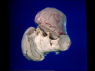
Bassett Collection of Stereoscopic Images of Human Anatomy
Exploration of the cerebellum and brain stem from above and to the right
Brachium pontis exposed
Image #27-5
KEYWORDS: Brain, Medulla, Midbrain, Pons.
Creative Commons
Stanford holds the copyright to the David L. Bassett anatomical images and has assigned Creative Commons license Attribution-Share Alike 4.0 International to all of the images.

For additional information regarding use and permissions, please contact the Medical History Center.

Exploration of the cerebellum and brain stem from above and to the right
Brachium pontis exposed
On the right side of the lingula, central lobule, quadrangular lobule and part of the superior semilunar lobule have been removed so that the brachium conjunctivum and brachium pontis are visible. The brain stem is sectioned at the junction of pons and mesencephalon.
- Horizontal cerebellar sulcus (sulcus intercruralis)
- Inferior semilunar lobe (crus II ansiform lobule)
- Medullary substance of posterior part quadrangular lobule (simplex lobule)
- Brachium conjunctivum (superior cerebellar peduncle)
- Brachium pontis (middle cerebellar peduncle)
- Superior semilunar lobe (crus I ansiform lobule)
- Posterior superior fissure
- Posterior part quadrangular lobule (simplex lobule)
- Primary fissure
- Anterior part quadrangular lobule (continuous with Culmen of monticulus)
- Medullary substance of culmen of right side
- Central lobule (midsagittal section)
- Decussation of superior cerebellar peduncle
- Pons
- Basilar artery
- Trigeminal nerve (V)