Bassett Collection of Stereoscopic Images of Human Anatomy
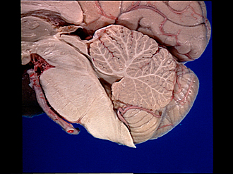
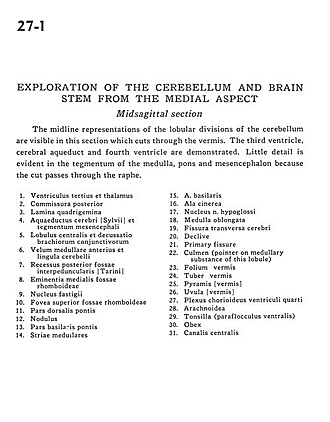
Exploration of the cerebellum and brain stem from the medial aspect
Midsagittal section
Image #27-1
KEYWORDS: Brain, Cerebellum, Medulla, Midbrain, Pons, Ventricules, Overview.
Creative Commons
Stanford holds the copyright to the David L. Bassett anatomical images and has assigned Creative Commons license Attribution-Share Alike 4.0 International to all of the images.

For additional information regarding use and permissions, please contact the Medical History Center.

Exploration of the cerebellum and brain stem from the medial aspect
Midsagittal section
The midline representations of the lobular divisions of the cerebellum are visible in this section which cuts through the vermis. The third ventricle, cerebral aqueduct and fourth ventricle are demonstrated. Little detail is evident in the tegmentum of the medulla, pons and mesencephalon because the cut passes through the raphe.
- Third ventricle and thalamus
- Posterior commissure
- Quadrigeminal plate
- Cerebral aqueduct and mesencephalic tegmentum
- Central lobule and decussation of superior cerebellar peduncle
- Anterior medullary velum and cerebellar lingula
- Posterior recess interpeduncular fossa
- Median eminence rhomboid fossa
- Fastigial nucleus
- Superior fovea rhomboid fossa
- Dorsal part of pons
- Nodulus
- Basilar part of pons
- Striae medullares
- Basilar artery
- Wing of gray matter
- Nucleus hypoglossal nerve
- Medulla oblongata
- Transverse cerebral fissure
- Declive
- Primary fissure
- Culmen (pointer on medullary substance of this lobule)
- Folium vermis
- Tuber vermis
- Pyramid (vermis)
- Uvula (vermis)
- Choroid plexus fourth ventricle
- Arachnoid
- Tonsil (ventral paraflocculus)
- Obex
- Central canal