
Bassett Collection of Stereoscopic Images of Human Anatomy
Exploration of the left half of the cerebellum and brain stem from its inferior and medial aspect
Lobulus gracilis viewed from below
Image #26-6
KEYWORDS: Brain, Cerebellum.
Creative Commons
Stanford holds the copyright to the David L. Bassett anatomical images and has assigned Creative Commons license Attribution-Share Alike 4.0 International to all of the images.

For additional information regarding use and permissions, please contact the Medical History Center.

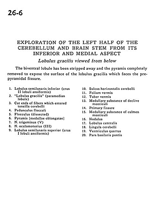
Exploration of the left half of the cerebellum and brain stem from its inferior and medial aspect
Lobulus gracilis viewed from below
The biventral lobule has been stripped away and the pyramis completely removed to expose the surface of the lobulus gracilis which faces the prepyramidal fissure.
- Inferior semilunar lobe (crus II ansiform lobule)
- Gracile lobule (paramedian lobule)
- Cut ends of fibers which entered cerebellar tonsil
- Floccular peduncle
- Flocculus (dissected)
- Pyramid (medulla oblongata)
- Trigeminal nerve (V)
- Oculomotor nerve (III)
- Superior semilunar lobe (crus I ansiform lobule)
- Horizontal cerebellar sulcus
- Folium vermis
- Tuber vermis
- Medullary substance of declive of cerebellum
- Primary fissure
- Medullary substance of Culmen of monticulus
- Nodulus
- Central lobule
- Cerebellar lingula
- Fourth ventricle
- Basilar part of pons