Bassett Collection of Stereoscopic Images of Human Anatomy
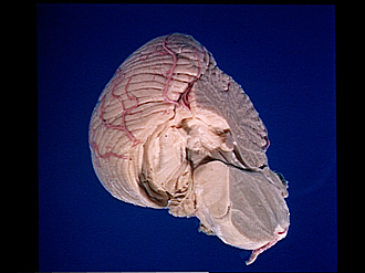
Exploration of the left half of the cerebellum and brain stem from its inferior and medial aspect
Tonsil removed; continuation of its medullary substance into pyramis
Image #26-4
KEYWORDS: Brain, Cerebellum, Medulla.
Creative Commons
Stanford holds the copyright to the David L. Bassett anatomical images and has assigned Creative Commons license Attribution-Share Alike 4.0 International to all of the images.

For additional information regarding use and permissions, please contact the Medical History Center.
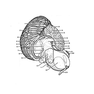
Exploration of the left half of the cerebellum and brain stem from its inferior and medial aspect
Tonsil removed; continuation of its medullary substance into pyramis
The lateral part of the tonsil has been removed. The biventral lobule is now exposed as it opposed the tonsil across the postpyramidal fissure (secondary fissure). Two major masses of fibers (5, 6) have been cut off as they entered the tonsil from the deep parts of the cerebellum. The more medial of these was previously followed into the uvula. The more lateral group of fibers is continuous medially into folia which join the pyramis.
- Inferior semilunar lobe (crus II ansiform lobule)
- Gracile lobule (paramedian lobule)
- Biventral lobule (dorsal paraflocculus) and postpyramidal fissure (secondary fissure)
- Branch of posterior inferior cerebellar artery (PICA)
- Cut ends of fibers which entered lateral part of cerebellar tonsil
- Cut ends of fibers which entered medial part of cerebellar tonsil
- Floccular peduncle
- Secondary flocculus
- Glossopharyngeal nerve (IX)
- Inferior olivary nucleus
- Pyramid (medulla oblongata)
- Basilar part of pons (midsagittal section)
- Folium vermis
- Primary fissure
- Pyramid (vermis)
- Cut ends of fibers which entered uvula
- Brachium conjunctivum (superior cerebellar peduncle)
- Nodulus
- Central lobule
- Cerebellar lingula
- Pineal body
- Quadrigeminal plate
- Occulomotor nerve (III)