Bassett Collection of Stereoscopic Images of Human Anatomy
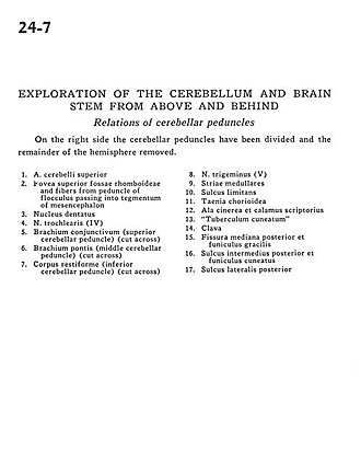
Exploration of the cerebellum and brain stem from above and behind
Relations of cerebellar peduncles
Image #24-7
KEYWORDS: Brain, Cerebellum.
Creative Commons
Stanford holds the copyright to the David L. Bassett anatomical images and has assigned Creative Commons license Attribution-Share Alike 4.0 International to all of the images.

For additional information regarding use and permissions, please contact the Medical History Center.
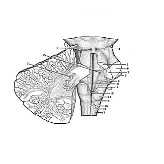
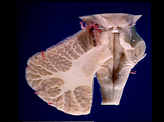
Exploration of the cerebellum and brain stem from above and behind
Relations of cerebellar peduncles
On the right side the cerebellar peduncles have been divided and the remainder of the hemisphere removed.
- Superior cerebellar artery
- Superior fovea rhomboid fossa and fibers from peduncle of flocculus passing into tegmentum of mesencephalon
- Dentate nucleus
- Trochlear nerve (IV)
- Brachium conjunctivum (superior cerebellar peduncle) (cut across)
- Brachium pontis (middle cerebellar peduncle) (cut across)
- Restiform body (inferior cerebellar peduncle) (cut across)
- Trigeminal nerve (V)
- Striae medullares
- Sulcus limitans
- Taenia chorioidea
- Wing of gray matter and calamus scriptorius
- Cuneate tubercle
- Clava
- Dorsal median sulcus and gracile fasciculus
- Dorsal intermediate sulcus and cuneate fasciculus
- Dorsal lateral sulcus