
Bassett Collection of Stereoscopic Images of Human Anatomy
Exploration of the cerebellum and brain stem from above and behind
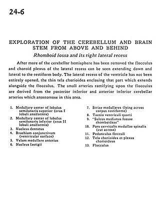
Rhomboid fossa and its right lateral recess
Image #24-6
KEYWORDS: Brain, Cerebellum, Ventricules.
Creative Commons
Stanford holds the copyright to the David L. Bassett anatomical images and has assigned Creative Commons license Attribution-Share Alike 4.0 International to all of the images.

For additional information regarding use and permissions, please contact the Medical History Center.

Exploration of the cerebellum and brain stem from above and behind
Rhomboid fossa and its right lateral recess
After more of the cerebellar hemisphere has been removed the flocculus and choroid plexus of the lateral recess can be seen extending down and lateral to the restiform body. The lateral recess of the ventricle has not been entirely opened, the thin tela chorioidea enclosing that part which extends alongside the flocculus. The small arteries ramifying upon the flocculus are derived from the posterior inferior and anterior inferior cerebellar arteries which anastomose in this area.
- Medullary center of superior semilunar lobe (crus I ansiform lobule)
- Medullary center of inferior semilunar lobe (crus II ansiform lobule)
- Dentate nucleus
- Brachium conjunctivum (superior cerebellar peduncle) (ventricular surface)
- Anterior medullary velum
- Fastigial nucleus
- Striae medullares (lying across restiform body (inferior cerebellar peduncle))
- Taenia fourth ventricle
- Median sulcus of rhomboid fossa
- Cervical part of medulla (cut across)
- Pedunculus flocculi
- Tela chorioidea and choroid plexus
- Flocculus