
Bassett Collection of Stereoscopic Images of Human Anatomy
Exploration of the cerebellum and brain stem from above and behind
Tela chorioidea and fourth ventricle; foramen of Magendie
Image #24-4
KEYWORDS: Brain, Cerebellum, Ventricules.
Creative Commons
Stanford holds the copyright to the David L. Bassett anatomical images and has assigned Creative Commons license Attribution-Share Alike 4.0 International to all of the images.

For additional information regarding use and permissions, please contact the Medical History Center.

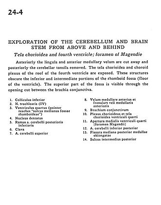
Exploration of the cerebellum and brain stem from above and behind
Tela chorioidea and fourth ventricle; foramen of Magendie
Anteriorly the lingula and anterior medullary velum are cut away and posteriorly the cerebellar tonsils removed. The tela chorioidea and choroid plexus of the roof of the fourth ventricle are exposed. These structures obscure the inferior and intermediate portions of the rhomboid fossa (floor of the ventricle). The superior part of the fossa is visible through the opening cut between the brachia conjunctiva.
- Inferior colliculus
- Trochlear nerve (IV)
- Fourth ventricle (pointer reaches "the median sulcus of the rhomboid fossae")
- Dentate nucleus
- Branch of posterior inferior cerebellar artery
- Clava
- Superior cerebellar artery
- Anterior medullary velum and frenulum
- Brachium conjunctivum (superior cerebellar peduncle)
- Choroid plexus and tela chorioidea fourth ventricle
- Medial aperture fourth ventricle (foramen of Magendie)
- Posterior inferior cerebellar artery
- Posterior median fissure of medulla oblongata
- Posterior intermediate sulcus