
Bassett Collection of Stereoscopic Images of Human Anatomy
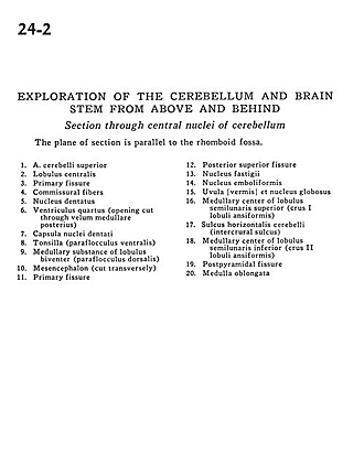
Exploration of the cerebellum and brain stem from above and behind
Section through central nuclei of cerebellum
Image #24-2
KEYWORDS: Brain, Cerebellum.
Creative Commons
Stanford holds the copyright to the David L. Bassett anatomical images and has assigned Creative Commons license Attribution-Share Alike 4.0 International to all of the images.

For additional information regarding use and permissions, please contact the Medical History Center.

Exploration of the cerebellum and brain stem from above and behind
Section through central nuclei of cerebellum
The plane of section is parallel to the rhomboid fossa.
- Superior cerebellar artery
- Central lobule
- Primary fissure
- Commissural fibers
- Dentate nucleus
- Fourth ventricle (opening cut through posterior medullary velum)
- Capsule of dentate nuclei
- Tonsil (ventral paraflocculus)
- Medullary substance of biventral lobule (dorsal paraflocculus)
- Mesencephalon (cut transversely)
- Primary fissure
- Posterior superior fissure
- Fastigial nucleus
- Emboliform nucleus
- Uvula (vermis) and globose nucleus
- Medullary center of superior semilunar lobe (crus l ansiform lobule)
- Horizontal cerebellar sulcus (intercrural sulcus)
- Medullary center of inferior semilunar lobule (crus II ansiform lobule)
- Postpyramidal fissure
- Medulla oblongata