Bassett Collection of Stereoscopic Images of Human Anatomy
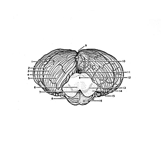
The Surfaces of the cerebellum and brain stem
Anterior aspect
Image #23-6
KEYWORDS: Brain, Cerebellum, Midbrain, Vasculature, Overview.
Creative Commons
Stanford holds the copyright to the David L. Bassett anatomical images and has assigned Creative Commons license Attribution-Share Alike 4.0 International to all of the images.

For additional information regarding use and permissions, please contact the Medical History Center.

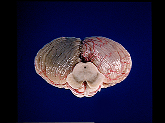
The Surfaces of the cerebellum and brain stem
Anterior aspect
The brain stem has been cut transversely through the mesencephalon. The right cerebellar hemisphere has been stripped of meninges and vessels to expose the pattern of cerebellar folia and sulci. The basilar artery has been removed, but the cut ends of pontine and mesencephalic branches as well as the superior cerebellar arteries have been left.
- Superior semilunar lobe (crus I ansiform lobule)
- Posterior superior fissure
- Posterior part quadrangular lobule (posterior semilunar lobule)
- Primary fissure
- Anterior part quadrangular lobule (anterior semilunar lobule)
- Red nucleus and brachium conjunctivum (superior cerebellar peduncle)
- Posterior recess interpeduncular fossa
- Pons
- Posterior cerebellar incisure
- Culmen of monticulus
- Anterior cerebellar incisure
- Cerebral aqueduct
- Substantia nigra
- Cerebral peduncle
- Horizontal cerebellar sulcus
- Superior cerebellar artery