Bassett Collection of Stereoscopic Images of Human Anatomy
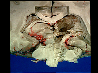
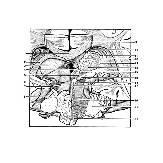
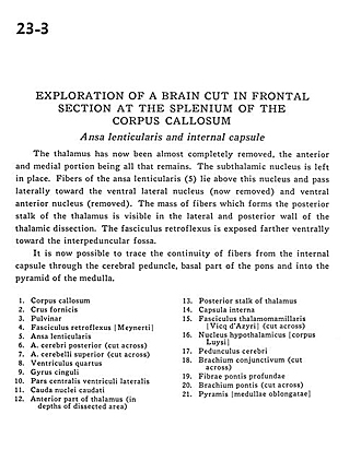
Exploration of a brain cut in frontal section at the splenium of the corpus callosum
Ansa lenticularis and internal capsule
Image #23-3
KEYWORDS: Brain, Diencephalon, Medulla, Midbrain, Pons.
Creative Commons
Stanford holds the copyright to the David L. Bassett anatomical images and has assigned Creative Commons license Attribution-Share Alike 4.0 International to all of the images.

For additional information regarding use and permissions, please contact the Medical History Center.
Exploration of a brain cut in frontal section at the splenium of the corpus callosum
Ansa lenticularis and internal capsule
The thalamus has now been almost completely removed, the anterior and medial portion being all that remains. The subthalamic nucleus is left in place. Fibers of the ansa lenticularis (5) lie above this nucleus and pass laterally toward the ventral lateral nucleus (now removed) and ventral anterior nucleus (removed). The mass of fibers which forms the posterior stalk of the thalamus is visible in the lateral and posterior wall of the thalamic dissection. The fasciculus retroflexus is exposed farther ventrally toward the interpeduncular fossa.
- Corpus callosum
- Fornix (crus)
- Pulvinar
- Retroflex fasciculus
- Ansa lenticularis
- Posterior cerebral artery (cut across)
- Superior cerebellar artery (cut across)
- Fourth ventricle
- Cingulate gyrus
- Central part lateral ventricle
- Caudate nucleus (tail)
- Anterior part of thalamus (in depths of dissected area)
- Posterior stalk of thalamus
- Internal capsule
- Mamillothalamic tract (cut across)
- Hypothalamic nucleus
- Cerebral peduncle
- Brachium conjunctivum (superior cerebellar peduncle) (cut across)
- Deep pontine fibers
- Brachium pontis (middle cerebellar peduncle) (cut across)
- Pyramid medulla oblongata