Bassett Collection of Stereoscopic Images of Human Anatomy
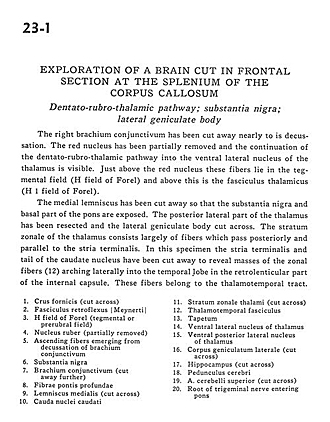
Exploration of a brain cut in frontal section at the splenium of the corpus callosum
Dentato-rubro-thalamic pathway; substantia nigra; lateral geniculate body
Image #23-1
KEYWORDS: Brain, Diencephalon, Midbrain, Pons, Telencephalon, Temporal lobe.
Creative Commons
Stanford holds the copyright to the David L. Bassett anatomical images and has assigned Creative Commons license Attribution-Share Alike 4.0 International to all of the images.

For additional information regarding use and permissions, please contact the Medical History Center.
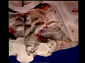
Exploration of a brain cut in frontal section at the splenium of the corpus callosum
Dentato-rubro-thalamic pathway; substantia nigra; lateral geniculate body
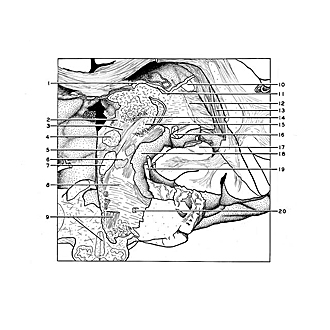
The right brachium conjunctivum has been cut away nearly to its decussation. The red nucleus has been partially removed and the continuation of the dentato-rubro-thalamic pathway into the ventral lateral nucleus of the thalamus is visible. Just above the red nucleus these fibers lie in the tegmental field (H field of Forel) and above this is the fasciculus thalamicus (H 1 field of Forel).
- Fornix (crus) (cut across)
- Retroflex fasciculus
- H field of Forel (tegmental or prerubral field)
- Red nucleus (partially removed)
- Ascending fibers emerging from decussation of brachium conjunctivum (superior cerebelIar peduncle)
- Substantia nigra
- Brachium conjunctivum (superior cerebellar peduncle) (cut away further)
- Deep pontine fibers
- Medial lemniscus (cut across)
- Caudate nucleus (tail)
- Stratum zonale thalami (cut across)
- Thalamotemporal fasciculus
- Tapetum
- Ventral lateral nucleus of thalamus
- Ventral posterior lateral nucleus of thalamus
- Lateral geniculate body (cut across)
- Hippocampus (cut across)
- Cerebral peduncle
- Superior cerebellar artery (cut across)
- Root of trigeminal nerve (V) entering pons