Bassett Collection of Stereoscopic Images of Human Anatomy
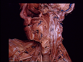
Dissection of anterior aspect of vertebral column
Cervical region.
Image #220-2
KEYWORDS: Cervical region, Muscles and tendons, Vertebral column.
Creative Commons
Stanford holds the copyright to the David L. Bassett anatomical images and has assigned Creative Commons license Attribution-Share Alike 4.0 International to all of the images.

For additional information regarding use and permissions, please contact the Medical History Center.
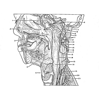

Dissection of anterior aspect of vertebral column
Cervical region.
The longus capitis muscle has been divided close to its origins (15) from the transverse processes of the third to the sixth cervical vertebrae and also at its insertion (1) into the occipital bone. The removal of the belly of this muscle has exposed the longus colli muscle (13) and the anterior arch of the atlas (9).
- Area of insertion of longus capitis muscle
- Nasal cavity
- Nasopharynx
- Oropharynx
- Larynx
- Upper pointer: Internal carotid artery (within carotid canal, accompanied by internal carotid plexus) Lower pointer: Jugular foramen (opened)
- Lateral rectus capitis muscle
- Upper pointer: Hypoglossal nerve (XII) Lower pointer: Anterior rectus capitis muscle
- Upper pointer: Anterior atlantooccipital membrane Lower pointer: Anterior arch of atlas (covered by connective tissue)
- Transverse process of atlas
- Lateral atlantoaxial joint (pointer on joint capsule)
- Ventral branch cervical nerve II
- Longus colli muscle
- Superior cervical ganglion
- Origins of longus capitis muscle
- Ventral branch cervical nerve III
- Anterior intertransverse muscle
- Sympathetic trunk
- Anterior longitudinal ligament (partially covered by prevertebral fascia)
- Middle cervical ganglion
- Transverse process of vertebra C. VII