
Bassett Collection of Stereoscopic Images of Human Anatomy
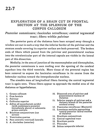
Exploration of a brain cut in frontal section at the splenium of the corpus callosum
Posterior commissure; fasciculus retroflexus; central tegmental tract; fibers within pulvinar
Image #22-7
KEYWORDS: Brain, Diencephalon, Midbrain.
Creative Commons
Stanford holds the copyright to the David L. Bassett anatomical images and has assigned Creative Commons license Attribution-Share Alike 4.0 International to all of the images.

For additional information regarding use and permissions, please contact the Medical History Center.
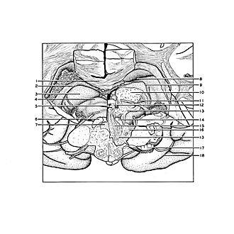
Exploration of a brain cut in frontal section at the splenium of the corpus callosum
Posterior commissure; fasciculus retroflexus; central tegmental tract; fibers within pulvinar
The posterior parts of the thalamus have been scraped away through a window cut out in such a way that the inferior border of the pulvinar and the stratum zonale covering its superior surface are both preserved. The broken ends of fibers which passed from the pulvinar and posterolateral nucleus into the retrolenticular part of the internal capsule are visible in the lateral part of this dissection.
- Corpus callosum
- Fornix (crus)
- Pulvinar
- Superior colliculus
- Cerebral aqueduct and entrance (with posterior commissure immediately above) and central gray matter
- Brachium conjunctivum (superior cerebellar peduncle) (cut across)
- Fourth ventricle
- Central part lateral ventricle
- Choroidal artery (anterior)
- Dissected area of pulvinar and posterolateral nucleus of thalamus
- Inferior margin of pulvinar and a thalamic branch of the posterior cerebral artery
- Retroflex fasciculus
- Medial lemniscus approaching ventral posterior lateral nucleus of thalamus
- Brachium conjunctivum (superior cerebellar peduncle)
- Central tegmental tract
- Brachium pontis (middle cerebellar peduncle)
- Flocculus
- Inferior temporal sulcus