
Bassett Collection of Stereoscopic Images of Human Anatomy
Exploration of a brain cut in frontal section at the splenium of the corpus callosum
Brachium conjunctivum and lateral lemniscus
Image #22-4
KEYWORDS: Brain, Midbrain.
Creative Commons
Stanford holds the copyright to the David L. Bassett anatomical images and has assigned Creative Commons license Attribution-Share Alike 4.0 International to all of the images.

For additional information regarding use and permissions, please contact the Medical History Center.

Exploration of a brain cut in frontal section at the splenium of the corpus callosum
Brachium conjunctivum and lateral lemniscus
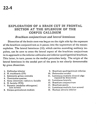
Dissection of the brain stem was begun on the right side by the exposure of the brachium conjunctivum as it passes into the tegmentum of the mesencephalon. The lateral lemniscus (12), which carries ascending auditory impulses, can be seen to cross the lateral aspect of the brachium conjunctivum in its approach to the inferior colliculus and inferior quadrigeminal brachium. This latter, in turn, passes to the medial geniculate body. The origin of the lateral lemniscus in the caudal part of the pons is not clearly demonstrable by gross dissection.
- Inferior colliculus
- Trochlear nerve (IV)
- Central gray matter
- Fourth ventricle
- Internal genu of roots of facial nerve
- Facial nerve (VII)
- Pyramid at medulla oblongata (cut across)
- Medial geniculate body
- Brachium of inferior colliculus
- Cerebral peduncle
- Medial lemniscus (lateral edge exposed by dissection)
- Lateral lemniscus
- Brachium conjunctivum (superior cerebellar peduncle)
- Brachium pontis (middle cerebellar peduncle)
- Medial lemniscus (cut across)
- Inferior olivary nucleus