Bassett Collection of Stereoscopic Images of Human Anatomy
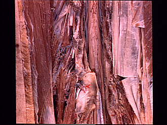
Lumbosacral meninges, spinal cord and nerve roots dissected from behind
Cauda equina, conus medullaris and filum terminale
Image #219-7
KEYWORDS: Central nervous system, Lumbar region, Sacral region, Vertebral column.
Creative Commons
Stanford holds the copyright to the David L. Bassett anatomical images and has assigned Creative Commons license Attribution-Share Alike 4.0 International to all of the images.

For additional information regarding use and permissions, please contact the Medical History Center.


Lumbosacral meninges, spinal cord and nerve roots dissected from behind
Cauda equina, conus medullaris and filum terminale
The lumbar part of the spinal cord has been exposed. The conus medullaris ends at the level of the arch of the second lumbar vertebra.
- Lateral cutaneous branch of dorsal branch thoracic nerve XII
- Medial intertransverse muscle
- Lamina (arch of vertebra) L. I (cut across)
- Lateral intertransversarius muscle
- Mamillary process vertebra L. II
- Dorsal branch lumbar nerve I
- Iliocostalis muscle
- Spinous process vertebra L. II (partly resected)
- Intervertebral joint capsule
- Dorsal roots thoracic nerve XII
- Denticulate ligament
- Intumescentia lumbalis
- Latissimus dorsi muscle (cut across)
- Cauda equina
- Longissimus thoracis muscle
- Superior articular surface
- Conus medullaris
- Filum terminale