Bassett Collection of Stereoscopic Images of Human Anatomy
Thoracic meninges, spinal cord and nerve roots dissected in relation to vertebral column
Transverse section of body of eighth thoracic vertebra illustrating relations of blood vessels, nerves and ligaments
Image #219-2
KEYWORDS: Central nervous system, Thoracic region, Vertebral column.
Creative Commons
Stanford holds the copyright to the David L. Bassett anatomical images and has assigned Creative Commons license Attribution-Share Alike 4.0 International to all of the images.
For additional information regarding use and permissions, please contact the Medical History Center.
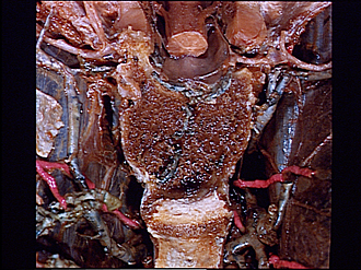
Thoracic meninges, spinal cord and nerve roots dissected in relation to vertebral column
Transverse section of body of eighth thoracic vertebra illustrating relations of blood vessels, nerves and ligaments
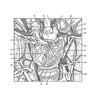
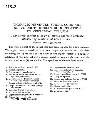
The thoracic part of the spinal cord has been exposed by a laminectomy. The upper thoracic vertebra have been completely removed for this view, including the upper half of the body of the eighth vertebra. The interrelations of the internal and external vertebral venous plexuses and the basivertebral vein (5) are visible. The specimen is viewed from above.
- Dorsal roots thoracic nerve IX
- Spinal cord
- Dura mater and arachnoid
- Pedicle (arch of vertebra) Th. VIIl (partially removed)
- Upper pointer: Anterior internal vertebral venous plexus Lower pointer: Basivertebral vein
- Body of vertebra Th. VIII (partly cut away)
- Ganglion of sympathetic trunk
- Greater splanchnic nerve
- Anterior longitudinal ligament
- Intervertebral disc Th. VII- VIII (remnant)
- Denticulate ligament
- Superior articular process vertebra Th. IX
- Dorsal branch thoracic nerve VIII
- Spinal ganglion
- Ventral branch thoracic nerve VIII
- Intervertebral foramen
- Posterior intercostal vein
- Parietal pleura
- Posterior intercostal artery VII
- Hemiazygos vein


