
Bassett Collection of Stereoscopic Images of Human Anatomy
Atlantooccipital joint, atlantoaxial joint and cervical vertebrae dissected from behind
Hypoglossal, accessory and vagus nerves; ventral rami of cervical nerves; internal carotid artery; internal jugular vein
Image #218-5
KEYWORDS: Cervical region, Vertebral column.
Creative Commons
Stanford holds the copyright to the David L. Bassett anatomical images and has assigned Creative Commons license Attribution-Share Alike 4.0 International to all of the images.

For additional information regarding use and permissions, please contact the Medical History Center.

Atlantooccipital joint, atlantoaxial joint and cervical vertebrae dissected from behind
Hypoglossal, accessory and vagus nerves; ventral rami of cervical nerves; internal carotid artery; internal jugular vein
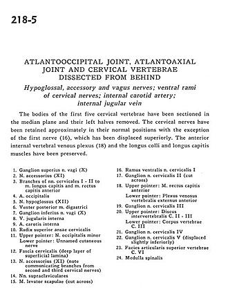
The bodies of the first five cervical vertebrae have been sectioned in the median plane and their left halves removed. The cervical nerves have been retained approximately in their normal positions with the exception of the first nerve (16), which has been displaced superiorly. The anterior internal vertebral venous plexus (18) and the longus colli and longus capitis muscles has been preserved.
- Superior ganglion vagus nerve (X)
- Accessory nerve (XI)
- Branches of cervical nerves I - II to longus capitis and anterior rectus capitis muscles
- Occipital artery
- Hypoglossal nerve (XII)
- Posterior belly digastric muscle
- Inferior ganglion vagus nerve (X)
- Internal jugular vein
- Internal carotid artery
- Superior root ansa cervicalis
- Upper pointer: Lesser occipital nerve Lower pointer: Unnamed cutaneous nerve
- Cervical fascia (deep layer of superficial lamina)
- Accessory nerve (XI) (note communicating branches from second and third cervical nerves)
- Supraclavicular nerves
- Levator scapulae muscle (cut across)
- Ventral branch cervical nerve I
- Ganglion cervical nerve II (cut across)
- Upper pointer: Anterior rectus capitis muscle Lower pointer: Anterior external vertebral venous plexus
- Ganglion of cervical nerve III
- Upper pointer: Intervertebral disc C. II- llI Lower pointer: Body of vertebra C. III
- Ganglion cervical nerve IV
- Ganglion cervical nerve V (displaced slightly inferiorly)
- Superior articular surface vertebra C. VI
- Spinal cord