Bassett Collection of Stereoscopic Images of Human Anatomy
Atlantooccipital joint, atlantoaxial joint and cervical vertebrae dissected from behind
Atlantooccipital and lateral atlantoaxial joints opened
Image #218-2
KEYWORDS: Cervical region, Vertebral column.
Creative Commons
Stanford holds the copyright to the David L. Bassett anatomical images and has assigned Creative Commons license Attribution-Share Alike 4.0 International to all of the images.

For additional information regarding use and permissions, please contact the Medical History Center.
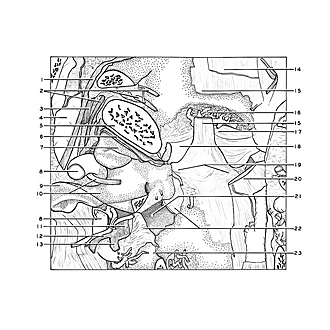
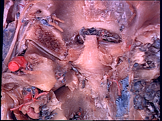
Atlantooccipital joint, atlantoaxial joint and cervical vertebrae dissected from behind
Atlantooccipital and lateral atlantoaxial joints opened
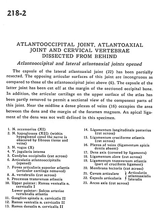
The capsule of the lateral atlantoaxial joint (22) has been partially resected. The opposing articular surfaces of this joint are incongruous as compared to those of the atlantooccipital joint above (6). The capsule of the latter joint has been cut off at the margin of the sectioned occipital bone. In addition, the articular cartilage on the upper surface of the atlas has been partly removed to permit a sectional view of the component parts of this joint. Near the midline a dense plexus of veins (16) occupies the area between the dens and the margin of the foramen magnum. An apical ligament of the dens was well defined in this specimen.
- Accessory nerve (XI)
- Hypoglossal nerve (XII) (within hypoglossal canal the nerve is obscured by fibrous tissue and veins)
- Vagus nerve (X)
- Internal jugular vein
- Occipital condyle (cut across)
- Atlantooccipital joint (opened)
- Superior articular facet of atlas (articular cartilage removed)
- Vertebral artery (cut across)
- Transverse process of atlas
- Upper pointer: Ventral branch cervical nerve I Lower pointer: Groove in atlas for vertebral artery
- Spinal ganglion cervical nerve II
- Ventral branch cervical nerve II
- Dorsal branch cervical nerve II
- Posterior longitudinal ligament (cut across)
- Cruciform ligament of atlas (cut across)
- Plexus of veins (ligamentum apicis dentis absent)
- Dens (axis) (covered by ligaments)
- Alar ligament (cut across)
- Transverse ligament of atlas (part of cruciform ligament)
- Tectorial membrane (cut across)
- Joint cavity
- Joint capsule lateralis (together 21 and 22 form lateral atlanto-occipital joint)
- Arch of axis (cut across)