Bassett Collection of Stereoscopic Images of Human Anatomy
Cervical meninges, spinal cord and nerve roots dissected from behind
General view of spinal cord in situ
Image #217-2
KEYWORDS: Central nervous system, Cervical region, Lumbar region, Sacral region, Vertebral column.
Creative Commons
Stanford holds the copyright to the David L. Bassett anatomical images and has assigned Creative Commons license Attribution-Share Alike 4.0 International to all of the images.

For additional information regarding use and permissions, please contact the Medical History Center.
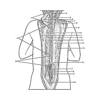
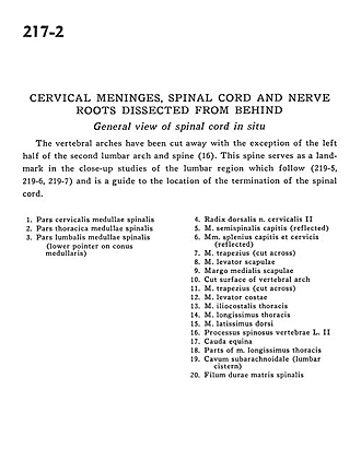
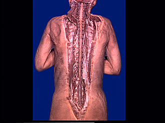
Cervical meninges, spinal cord and nerve roots dissected from behind
General view of spinal cord in situ
The vertebral arches have been cut away with the exception of the left half of the second lumbar arch and spine (16). This spine serves as a landmark in the close-up studies of the lumbar region which follow (219-5,219-6,219-7)and is a guide to the location of the termination of the spinal cord.
- Cervical part spinal cord
- Spinal cord thoracic part
- Spinal cord lumbar part (lower pointer on conus medullaris)
- Dorsal roots cervical nerve II
- Semispinalis capitis muscle (reflected)
- Splenius capitis and cervicis muscles (reflected)
- Trapezius muscle (cut across)
- Levator scapulae muscle
- Medial margin of scapula
- Cut surface of vertebral arch
- Trapezius muscle (cut across)
- Levator costarum muscle
- Iliocostalis muscle
- Longissimus thoracis muscle
- Latissimus dorsi muscle
- Spinous process vertebra L. II
- Cauda equina
- Parts of longissimus thoracis muscle
- Subarachnoid space (lumbar cistern)
- Filum terminale