
Bassett Collection of Stereoscopic Images of Human Anatomy
Ligaments and joints of lumbosacral spine and pelvic girdle
Intervertebral articulations viewed from above and behind
Image #215-5
KEYWORDS: Lumbar region, Muscles and tendons, Sacral region, Thoracic region, Vertebral column.
Creative Commons
Stanford holds the copyright to the David L. Bassett anatomical images and has assigned Creative Commons license Attribution-Share Alike 4.0 International to all of the images.

For additional information regarding use and permissions, please contact the Medical History Center.

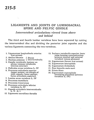
Ligaments and joints of lumbosacral spine and pelvic girdle
Intervertebral articulations viewed from above and behind
The third and fourth lumbar vertebrae have been separated by cutting the intervertebral disc and dividing the posterior joint capsules and the various ligaments connecting the two vertebrae.
- Anterior longitudinal ligament (cut across)
- Anulus fibrosus
- Nucleus pulposus (both 2 and 3 are part of the intervertebral disc)
- Vertebral canal (pointer on anterior internal vertebral venous plexus)
- Pedicle (arch of vertebra) L. IV
- Superior articular process (upper pointer, cut margin of joint capsule; lower pointer, superior articular surface)
- Lamina (arch of vertebra) L. IV
- Mamillary process
- Transverse process
- Inferior articular process vertebra L. IV
- Intervertebral joint capsule L. V - S. 1
- Dorsal sacroiliac ligament
- Superior vertebral incisure (note veins forming communications between internal and external vertebral venous plexuses)
- Ligamentum flavum (cut across)
- Interspinous ligament
- Iliolumbar ligament
- Lateral part sacrum
- Iliac crest
- Supraspinous ligament (overlying spinous process vertebra L. IV)