
Bassett Collection of Stereoscopic Images of Human Anatomy
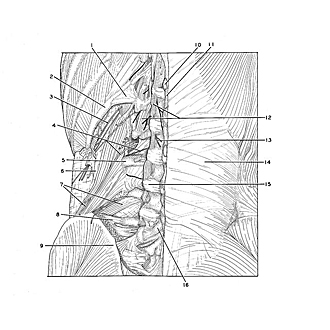
Dissection of thoracic and lumbosacral regions of back from a posterior approach
Quadratus lumborum muscle, posterior view
Image #214-6
KEYWORDS: Lumbar region, Muscles and tendons, Sacral region, Thoracic region, Vertebral column.
Creative Commons
Stanford holds the copyright to the David L. Bassett anatomical images and has assigned Creative Commons license Attribution-Share Alike 4.0 International to all of the images.

For additional information regarding use and permissions, please contact the Medical History Center.

Dissection of thoracic and lumbosacral regions of back from a posterior approach
Quadratus lumborum muscle, posterior view
The middle layer of thoracolumbar fascia has been removed from the posterior surface of the quadratus lumborum muscle (7). Small slips of origin join the quadratus lumborum from the transverse processes of the second and third lumbar vertebrae.
- Lumbocostal ligament
- Rib XII
- Subcostal nerve
- Left pointer: Dorsal branch lumbar artery II Right pointer: Dorsal branch lumbar nerve II
- Transverse process of vertebra L. III
- Thoracolumbar fascia (anterior layer)
- Quadratus lumborum muscle
- Iliolumbar ligament
- Posterior superior iliac spine
- Spinous process vertebra Th. XII
- Supraspinous ligament
- Intertransverse muscles
- Dorsal branch lumbar nerve II
- Erector spinae muscle (covered by thoracolumbar fascia)
- Intervertebral joint capsule L. III-IV
- Lamina (arch of vertebra) L. V